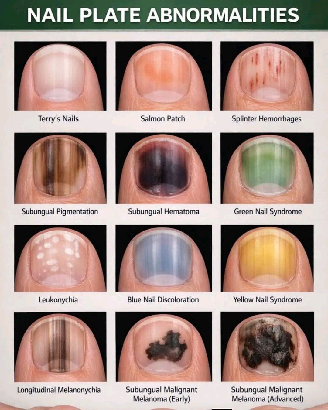
1. Terry’s Nails
→ Nail plate appears diffusely white or pale with a narrow distal pink or brown band
→ Due to decreased vascularity of nail bed
→ Common associations
→ Hypoalbuminemia
→ Liver cirrhosis
→ Congestive heart failure
→ Diabetes mellitus
→ Chronic kidney disease
2. Salmon Patch (Oil Drop Sign)
→ Yellow-pink translucent patch visible beneath nail plate
→ Represents psoriatic involvement of nail bed
→ Highly specific for nail psoriasis
→ Often associated with
→ Pitting
→ Onycholysis
→ Subungual hyperkeratosis
3. Splinter Hemorrhages
→ Thin, longitudinal reddish-brown streaks under nail plate
→ Caused by rupture of nail bed capillaries
→ Seen in
→ Trauma (most common)
→ Infective endocarditis
→ Vasculitis
→ Antiphospholipid syndrome
4. Subungual Pigmentation (Low-Grade Hematoma)
→ Brownish or reddish discoloration beneath nail plate
→ Usually due to minor repeated trauma
→ Pigment does not involve nail matrix
→ Lesion grows out with nail growth
→ Important differential: melanoma
5. Subungual Hematoma
→ Dark red, maroon, or black discoloration
→ Represents acute bleeding under nail plate
→ Usually painful initially
→ Common after blunt trauma
→ Color migrates distally with nail growth
6. Green Nail Syndrome
→ Green-black discoloration of nail plate
→ Caused by Pseudomonas aeruginosa infection
→ Occurs in moist environments
→ Seen in
→ Onycholysis
→ Chronic water exposure
→ Artificial nails
7. Leukonychia
→ White spots or transverse white streaks on nail plate
→ Due to abnormal keratinization of nail matrix
→ Most commonly from minor trauma
→ Lesions move distally with nail growth
→ Not due to calcium deficiency
8. Blue Nail Discoloration
→ Diffuse bluish coloration of nail plate
→ Caused by reduced oxygenation or altered hemoglobin
→ Seen in
→ Cyanosis
→ Methemoglobinemia
→ Drug-induced (e.g., antimalarials, amiodarone)
→ Advanced systemic illness
9. Yellow Nail Syndrome
→ Yellow, thickened, slow-growing nails
→ Loss of cuticle often seen
→ Classic triad
→ Yellow nails
→ Lymphedema
→ Respiratory disease (chronic sinusitis, bronchiectasis, pleural effusion)
10. Longitudinal Melanonychia
→ Single or multiple brown-black vertical bands
→ Pigment originates from nail matrix melanocytes
→ Common causes
→ Benign melanocytic activation
→ Junctional nevus
→ Physiological (especially in darker skin types)
→ Requires differentiation from melanoma
11. Subungual Malignant Melanoma (Early)
→ Irregular pigmented band with
→ Uneven borders
→ Color variation
→ Widening proximally
→ Hutchinson sign may be present
→ High suspicion if single digit involved
→ Does not move with nail growth
12. Subungual Malignant Melanoma (Advanced)
→ Large irregular pigmented mass
→ Nail plate destruction or lifting
→ Possible ulceration or bleeding
→ Represents late presentation
→ Poor prognosis if diagnosis delayed
Medical disclaimer: Medinaz Academy does not provide medical advice.
